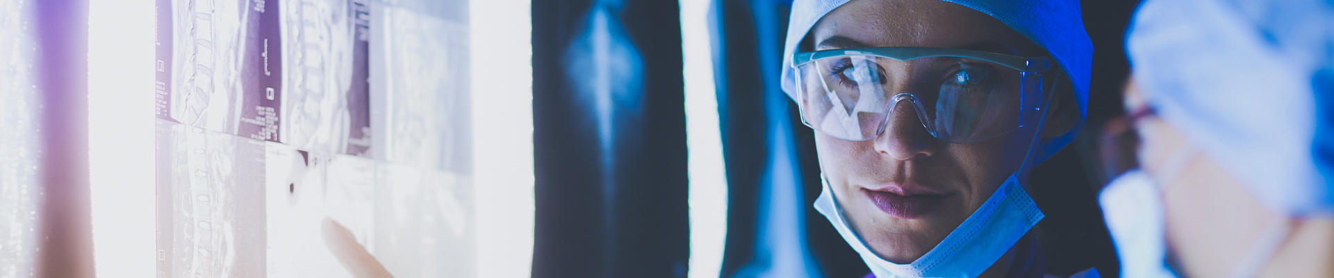
Imaging
Our Imaging Services Department provides vital diagnostic images to the physician and care team. These complex and extremely detailed images are essential in helping physicians and providers identify a patient's injury or illness. The greater the detail, the more accurate the diagnosis. The Imaging Services Department is available to patients 24 hours a day, every day. Board Certified Physicians and licensed, certified staff provide services using advanced diagnostic technology from the finest imaging manufacturers in the world. Service is provided in a relaxed, comfortable setting. Patients and family members will be treated with respect, dignity and kindness.The Imaging Services Department at Hampton Regional is fully digital, allowing patient's diagnostic images to be sent via high speed, secure computer connections to physicians and hospitals anywhere. Equipment and imaging services available at the hospital include:
Bone Density Scan
Digital Radiography
Ultrasound
Computed Tomography (CT Scan)
Mammography
MRI
Echocardiography
Nuclear Medicine
ABOUT THE RADIOLOGISTS
Hampton Regional Medical Center, in affiliation with MUSC Health, is dedicated to providing top-tier, local imaging services led by experts in their fields. Our radiology team is overseen by board-certified MUSC Radiologist Dr. Brittany Dobson, a specialist in breast health.Dr. Dobson brings her specialized expertise in breast imaging to our community, ensuring that patients at Hampton Regional Medical Center receive expert care close to home. Our facility is equipped with state-of-the-art technology, including a 3D mammography program, which allows for advanced diagnostics and early detection of breast health concerns, all under the expert guidance of Dr. Dobson and her team.
Digital Radiography
General-purpose radiology, previously called X-ray, is simply a procedure used to evaluate injury to the extremities: arms, legs, hands, and feet. This type of imaging is frequently used to diagnose fractures or broken bones. Hampton Regional's T.Rad Plus uses the most advanced imaging technology available.Ultrasound:
Ultrasound, or sonography, involves using high-frequency sound waves to produce pictures of the inside of the body. Ultrasound images are captured in real-time and show the structure and movement of the body's internal organs, as well as blood flowing through blood vessels. This procedure is used to examine internal organs such as the heart and blood vessels, liver, gallbladder, pancreas, kidneys, bladder, uterus, ovaries, thyroid, and fetus in pregnant patients. Ultrasound is helpful in diagnosing a variety of conditions.Computed Tomography (CT Scan):

Computed tomography (CT) scanning blends the traditional use of X-rays with the latest computer innovations. CT imaging uses special x-ray equipment to produce cross-sectional images or pictures of the inside of the body, and a computer reconstructs these slices to produce a 3D image of the areas being studied. When the image slices are reassembled by computer software, the result is a very detailed multidimensional view of the body's interior. CT scans of internal organs, bone, soft tissue and blood vessels provide greater clarity than conventional x-ray exams. A CT scan is a quick and painless procedure. It allows the technologist to acquire images in just a few seconds while you lie on the patient table. CT imaging can also play a significant role in the detection, diagnosis, and treatment of vascular disorders that can lead to stroke, gangrene or kidney failure.
Digital Mammography:
 Constance Ginn – Radiologic Technologist
Mammograms are used as a screening tool to detect early breast cancer in women. Digital Mammography is a specific type of imaging in which the x-ray films are replaced by solid-state detectors that convert x-rays into electrical signals. The electrical signals are used to produce images of the breast that can be seen on a computer. Digital mammography is proven to be more effective than conventional mammography.
MRI:
 Adam Petrus – MRI Technician
MRI (Magnetic Resonance Imaging) uses a powerful magnetic field and a computer to produce detailed pictures of organs, soft tissues, bone, and other internal body structures. This is a non-invasive, usually painless medical test that does not use radiation. Physicians use the MRI examination to help diagnose or monitor treatment for tumors of the chest, abdomen, or pelvis; lesions of the liver or other internal organs; and tumors or abnormalities of the reproductive organs.